FIG. 3.
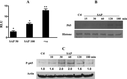
SAP induces NF-κB transactivation and p65 phosphorylation. (A) Caco-2 cells were transfected with pNF-κB-Luc plasmid with or without the positive control (p-MEKK), treated with different concentrations of SAP for 6 h, and assayed for luciferase activity as described in the text. Results are from three independent studies. *, P < 0.05; **, P < 0.01; RLU, relative luciferase units compared to values for the untreated cells; +ve, positive control. (B) Cells were treated with 100 μg/ml of SAP for different time periods, nuclear extracts were prepared as described in the text, and 10 μg/ml of protein was separated by SDS-PAGE and checked for p65 nuclear translocation. The blot was stripped and reprobed with histone antibody for normalization. A blot from one of three experiments is shown. Ctl, control. (C) Whole-cell lysates from cells treated as described above were subjected to Western blotting and probed with antibody specific to phosphorylated p65 (P-p65) and then with antibody against actin. Representative blots from two experiments, in which P-p65 levels are normalized to actin following densitometric scanning of blots, are shown. Expression in control cells was assigned an arbitrary value of 1, and expressions relative to this value are shown for treated cells.

