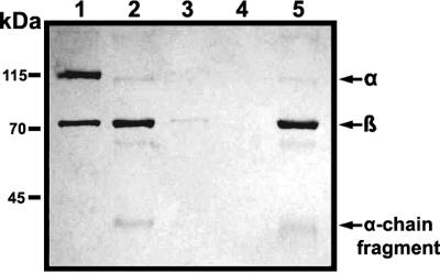
FIG. 5.
Effect of GelE on cell surface deposition of C3. The protein samples on E. faecalis GM were detected via immunoblot analysis. The nitrocellulose membrane was probed with anti-C3 antibody, which had been diluted to a concentration of 1:1,000 in antibody solution (1% skim milk in Tris-buffered saline containing 0.05% Tween 20). The secondary antibody used in this experiment was horseradish peroxidase-conjugated goat anti-rabbit antibody, as appropriate, at a dilution of 1:8,000. One microgram of purified C3 was employed as a control. Lane 1, purified C3; lane 2, NHS incubated with PBS; lane 3, hNHS incubated with PBS; lane 4, NHS incubated with GelE; lane 5, NHS incubated with SprE. Arrows indicate the α chain, β chain, and α-chain fragments of human C3.

