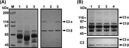
FIG. 6.
Degradation of complement factor C3 α chain by GelE, as determined via SDS-PAGE. (A) Proteolysis of the C3 α chain in NHS by GelE. Left panel, 8% SDS-PAGE gel conducted with NHS; right panel, immunoblot analysis. Lane M, molecular mass marker; lane 1, NHS incubated with PBS; lane 2, NHS incubated with GelE; lane 3, NHS incubated with SprE. Arrows indicate the intact α and β chains of C3 present in NHS. (B) Change in the C3 α chain in NHS by E. faecalis GM. Equal volumes of the mixtures were removed and subjected to SDS-PAGE and immunoblot analysis. Upper panel, an SDS-PAGE gel; lower panel, immunoblot analysis. Lane 1, NHS incubated with PBS; lane 2, NHS incubated with E. faecalis GM for 6 h; lane 3, NHS incubated with E. faecalis GM for 12 h; lane 4, NHS incubated with E. faecalis GM for 24 h.

