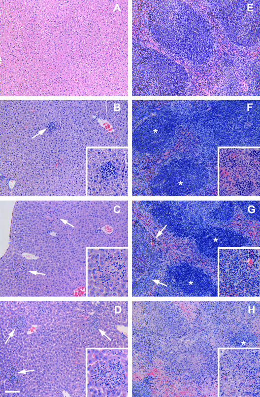
FIG. 4.
Histopathological findings of the livers (A to D) and spleens (E to H) of mice killed at 0, 1, 2, and 3 days after inoculation by gavage with 108 CFU of type A F. tularensis strain FSC033. The liver (A) and spleen (E) from a mouse killed at dpi 0 showed normal histological appearance. Mild, focal monocytic, and neutrophilic infiltration (arrow) was observed in the liver (B) and spleen (F) of a mouse killed at dpi 1. (C) At dpi 2, marked inflammatory infiltration and necrosis of individual hepatocytes (arrows) was observed in the liver. (G) The spleen showed moderate infiltration of neutrophils in the red pulp regions (arrows) as well as within some lymphoid follicles. (D and H) At dpi 3, the liver showed multiple foci and areas of inflammation (arrows) (D), and the spleen showed the severe destruction of lymphoid tissues and neutrophil infiltration, with only the remnants of normal lymphoid follicles (*) evident in some severely affected areas (H). *, Lymphoid follicles. Hematoxylin and eosin staining was used. Bar, 40 μm for the main images and 20 μm for all of the inset images.

