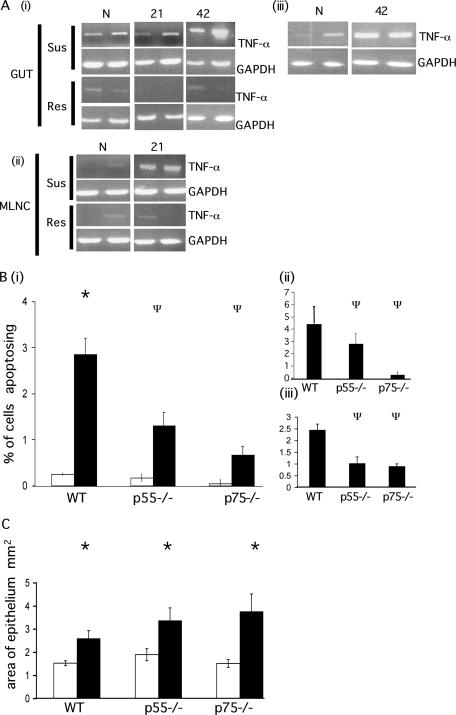
FIG. 3.
Apoptosis in TNFR-deficient animals during chronic T. muris infection. (A) The expression of TNF-α in the gut (i) and MLNC (ii) of AKR (susceptible) and BALB/c (resistant) mice was assessed by PCR at various times p.i. (iii) TNF-α expression in the gut tissue of C57BL/6 mice following low-dose infection was assessed by PCR. GAPDH (glyceraldehyde-3-phosphate dehydrogenase) levels were assessed as a control transcript. N, naive animals; 21, 21 days p.i.; 42, 42 days p.i. (B) p55−/−, p75−/−, and WT mice were infected with T. muris. The levels of apoptosis were assessed at day 42 p.i. and are expressed as the total percentage of cells apoptosing (i) and as the percentage of cells apoptosing in the base of the crypt (positions 1 to 4) (ii) and higher up the crypt axis (positions 11 to 30) (iii). Bars: □, naive animals; ▪, infected animals. (C) The area of the epithelium was assessed at day 42 p.i. Bars: □, naive animals; ▪, infected animals. Each datum point represents the mean of four animals ± the SEM. *, ANOVA and Tukey tests showed a statistically significant increase in infected versus naive animals (P < 0.001); Ψ, ANOVA and Tukey tests showed a statistically significant reduction in gene-deficient mice compared to WT mice (P < 0.001).

