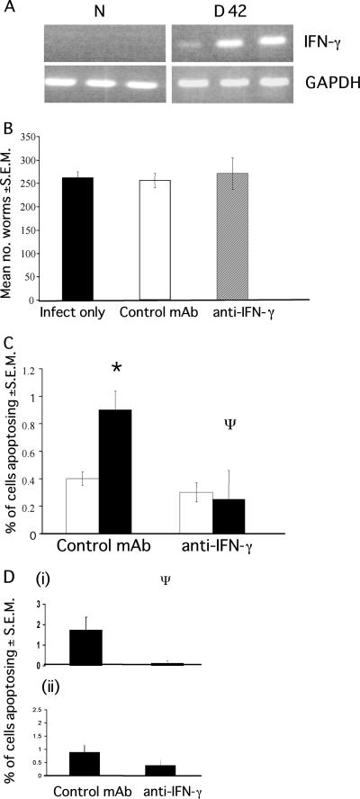
FIG. 4.
Apoptosis levels in SCID mice after neutralization of IFN-γ. SCID mice received either anti-IFN-γ (XMG1.6) or a control MAb GL113 at 1 mg/injection every 4 days for the duration of infection. (A) IFN-γ is expressed in the cecum during low-dose infection in C57BL/6 mice (B) Worm recovery from SCID mice treated with either anti-IFN-γ or control MAb. Each value represents a mean of four animals ± the SEM. (C) The levels of apoptosis were assessed at day 42 p.i. (□, naive mice; ▪, infected mice). (D) The position of apoptosing cells in the ceca of infected animals was assessed in the base of the crypt (cell positions 1 to 4) (i) and higher up the crypt (cell positions 11 to 15) (ii). Each value represents the mean of four animals ± the SEM. *, ANOVA and Tukey tests revealed a statistically significant difference between naive and infected animals and between control and anti-IFN-γ-treated mice (P < 0.01).

