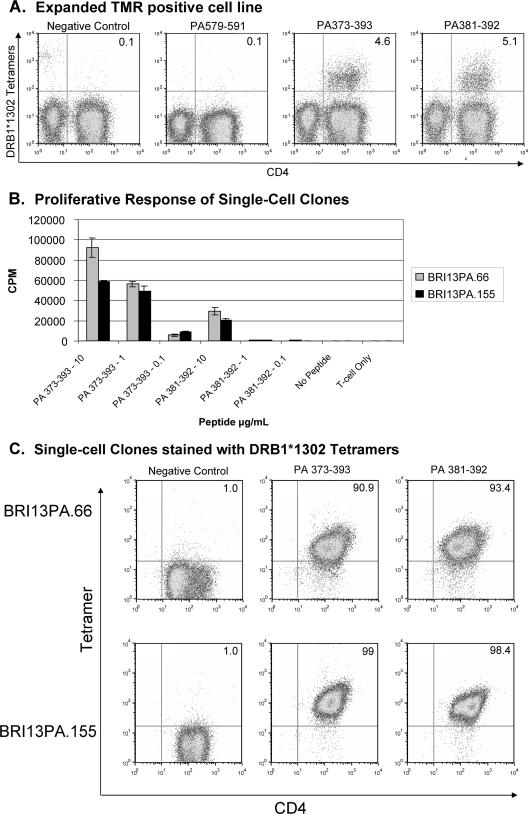
FIG. 3.
Characterization of the DRB1*1302 PA-positive tetramer population, BRI13PA cells, by tetramer staining and proliferation assay. (A) Tetramer staining of the bulk-sorted DRB1*1302 PA 381-392 sample after expansion with mitogen stimulation. The DRB1*1302 tetramers individually loaded with mortalin10A (Negative Control peptide) or PA peptides were incubated with cells for 2.5 h at 37°C, followed by CD4 staining for 30 min on ice. (B) BRI13PA clones proliferated to specific PA peptides in the presence of irradiated DRB1*1302 PBMC. [3H]thymidine was added 48 h after antigen and detected by scintillation counting 20 to 24 h later. Greater proliferation to the longer PA 373-393 peptide sequence correlates with the relative strength of peptide binding. (C) BRI13PA clones were stained with a negative control tetramer (DRB1*1302 mortalin10A; peptide sequence, AIKGAVVGIALG), DRB1*1302 PA 373-393, and DRB1*1302 PA 381-392 and analyzed by flow cytometry. The flow cytometry plots show cells in the live lymphocyte gate.

