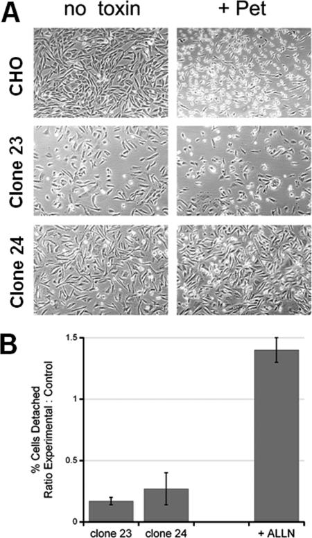
FIG. 5.
ERAD dysfunction blocks Pet intoxication. (A) Wild-type CHO cells and two mutant CHO cell lines with ERAD dysfunction (clones 23 and 24) were incubated for 10 h in the absence or presence of 40 μg Pet/ml. Images were taken at a magnification of ×10. (B) Wild-type CHO cells, mutant clone 23, mutant clone 24, and wild-type CHO cells treated with 10 μM of the proteasome inhibitor ALLN were exposed to 40 μg Pet/ml for 20 h. The percentage of detached cells was then determined for each condition. The results are expressed as the ratio of the experimental value to the control value, where the experimental value is the percentage of detached cells from the mutant cell line or ALLN-treated cells and the control value is the percentage of detached cells from the wild-type CHO cells. The averages ± standard deviations of three (mutant cell lines) or five (ALLN treatment) independent experiments are shown.

