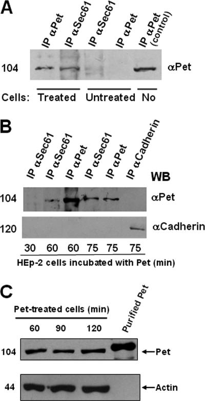
FIG. 7.
Pet and Sec61p interaction and full-length Pet translocation. (A and B) Coimmunoprecipitation of Pet and the Sec61p translocon. (A) Coimmunoprecipitation of Pet by using antibodies against Sec61α or Pet in cells treated with Pet for 1 h or in untreated cells. IP, immunoprecipitation. (B) Coimmunoprecipitation at various times. HEp-2 cells incubated with 37 μg Pet/ml for 30, 60, or 75 min were lysed, and the resulting supernatants were immunoprecipitated with either anti-Sec61α, anti-Pet, or anti-cadherin antibodies. A Western blot analysis of the immunoprecipitated proteins was conducted with anti-Pet antibodies, followed by a secondary peroxidase-labeled antibody. The position of a molecular weight marker is indicated on the left. (C) Pet detection in cytoplasmic fractions from Pet-treated cells. HEp-2 cells incubated with 37 μg Pet/ml for 60, 90, or 120 min were lysed and ultracentrifuged, and soluble cytoplasmic fractions were obtained. Equivalent volumes of the samples were subjected to SDS-PAGE, transferred to nitrocellulose membranes, and probed with a rabbit anti-Pet polyclonal antibody (top). Protein loading was monitored by stripping and reprobing with a mouse monoclonal anti-actin antibody (bottom).

