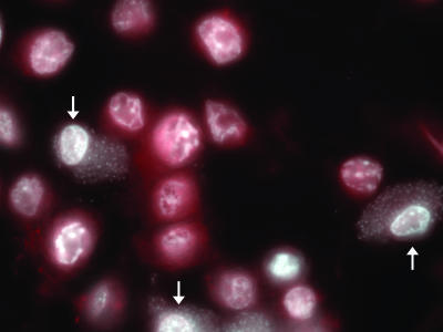
FIG. 2.
Activated caspase 3 is not present in Shigella-infected cells after STS treatment. Immunofluorescence analysis of an infected monolayer stained for activated caspase 3 (red) in addition to DAPI staining (white) after STS treatment in the modified assay. The arrows point to infected cells. This image is representative of cells observed in three independent experiments.

