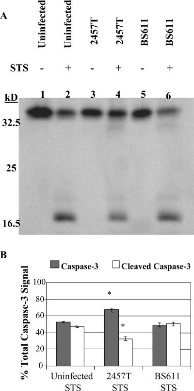
FIG. 3.
Western blot analysis for the activation of caspase 3. (A) Whole-cell lysates were separated by SDS-PAGE and analyzed by immunoblotting with an anti-caspase 3 antibody, which recognizes the full-length, inactive form of caspase 3 (35 kDa) and the large fragment resulting from cleavage during activation (17 kDa). Lane 1, uninfected HeLa cells; lane 2, uninfected HeLa cells with STS treatment; lane 3, 2457T-infected cells; lane 4, 2457T-infected cells with STS treatment; lane 5, BS611-infected cells; lane 6, BS611-infected cells with STS treatment. (B) Results of densitometry analysis of the percentage of the total caspase 3 detected in the Western blot are divided into the inactive form (caspase 3) and the activated form (cleaved caspase 3). The average percentage of each form is shown, with error bars representing the standard deviations from three independent experiments. *, P < 0.001 using Tukey's analysis of variance post hoc test, showing a significant difference between the 2457T STS treatment group and the other two treatment groups.

