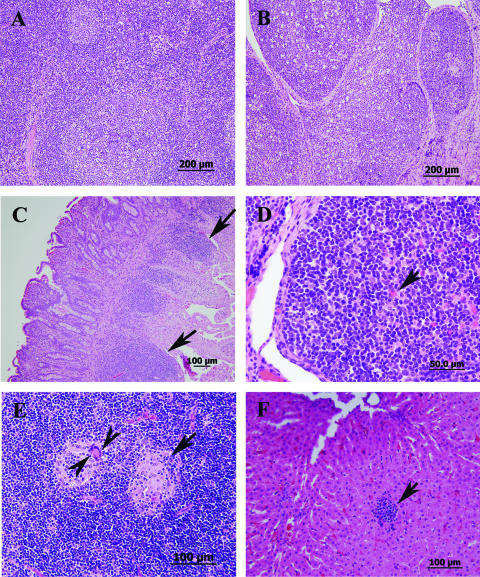
FIG. 3.
Histopathology of calf tissues at different times following infection with M. avium subsp. paratuberculosis strain K-10. All sections were stained with hematoxylin and eosin. (A and B) Sections of a normal lymph node (A) and Peyer's patches of the ileum (B) from a control calf sacrificed 60 days after infection. (C) Small intestine with enlarged lamina propria (arrows) at 8 months postinfection. (D) Ileal Peyer's patches with large follicles and Mott cells (arrows) at 9 months postinfection. (E) Mesenteric lymph node with necrotic centers (arrow), scattered macrophages, and giant cells (arrowheads) at 9 months postinfection. (F) Liver with macrophage aggregate (arrow) at 9 months postinfection.

