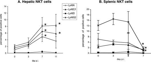
FIG. 3.
Kinetics of hepatic and splenic Ly49+ NKT cells during primary P. yoelii infection in C57BL/6 mice. Percentages of Ly49+ cells among hepatic (A) and splenic (B) NKT cells in noninfected (day 0) B6 mice and at different time points after injection of 4,000 sporozoites are shown. Cells were stained with anti-NK1.1 and anti-CD3 mAbs and anti-Ly49A, -Ly49C/I, -Ly49D, or -Ly49G2 mAbs. The percentages of Ly49+ cells were determined by flow cytometry analysis. Data are representative of three different experiments with three mice per time point. Results are expressed as mean values ± SD. *, the P value was <0.05 between control (day 0) and infected animals.

