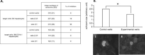
FIG. 7.
In vitro inhibition assay of P. yoelii liver-stage development by parasite-activated hepatic DN NKT cells. (A) Sorted hepatic DN NK1.1+ CD5+ cells isolated from infected B6 mice at day 10 p.i. were added to primary cultures of B6 or B6.CD1d−/− hepatocytes at two different NKT cell/hepatocyte ratios (2.5/1 and 5/1) 3 h after sporozoite addition. Forty-five hours later, schizonts were quantified in control (only medium added) and experimental (DN NKT cells added) wells, and the mean of duplicate wells was calculated. Data representative of two independent and three independent experiments performed with B6 and B6.CD1d−/− hepatocytes, respectively, are shown. (B) Mean diameters ± SD of the intrahepatic schizonts in control and experimental wells of B6 hepatocyte cultures. *, the P value of <0.001. Representative pictures show schizonts found in control (i) and experimental (ii) wells (magnification, ×400).

