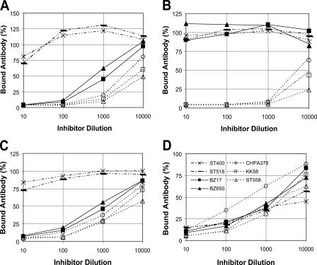
FIG. 1.
Antibody bound to ELISA plates (y axis) against the dilution of pneumococcal lysates (x axis). Lysates include two 6Aβ isolates (solid symbols with continuous lines), three 6Aα isolates (open symbols with dotted lines), and two 6B isolates (dashed connecting lines). Antibodies used for the assay were Hyp6AG1 (A), Hyp6AM3 (B), rabbit serum pool Q (C), and rabbit “factor 6b” serum (D).

