FIG. 1.
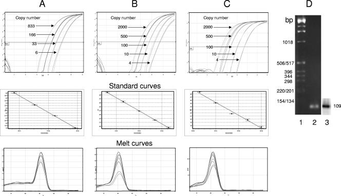
cVL and iVL can be quantitated by real-time PCR. (A) β-Globin DNA was quantitated from total DNA extracted from a mix of three chronically infected cell lines, as used for HIV standards, and subjected to real-time PCR quantitation as described in Materials and Methods. Real-time amplification curves (cycle number versus normalized florescence), derived standard curves (concentration versus CT) and melt analysis (oC versus derivative of fluorescence/derivative of time) of PCR products are shown. (B) Cell-associated HIV DNA was quantitated from total DNA extracted from a mix of three chronically infected cell lines, diluted in a background of uninfected DNA from 10,000 cell equivalents, and subjected to real-time PCR quantitation of a region of the HIV LTR as described in Materials and Methods. Real-time amplification curves, derived standard curves and melt analysis of PCR products are shown. (C) Integrated HIV DNA was quantitated from total DNA as described for panel A and diluted in a background of uninfected DNA from 50,000 cell equivalents. DNA was amplified in a first-round PCR with Alu and PBS-659 primers followed by dilution and second-round PCR amplification of the HIV LTR, as described for panel A and in Materials and Methods. Real-time amplification curves, derived standard curves, and melt analysis of PCR products are shown. (D) Gel analysis of PCR products. Second-round PCR products were subjected to agarose gel electrophoresis, stained with ethidium bromide, and photographed. Lane 1, molecular weight markers; lane 2, PCR product. DNA was then subjected to Southern analysis using an HIV-specific LTR probe and analyzed by autoradiography (lane 3).

