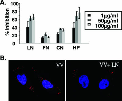
FIG. 1.
Laminin blocks IMV binding to BSC40 cells. (A) Different concentrations of soluble LN, FN, CN, or HP were mixed with purified IMV (150 PFU) for 30 min at 4°C and then added to cells for 30 min at 4°C, and the percent inhibition of virus binding to cells was determined by plaque assay as described in Materials and Methods. (B) Confocal immunofluorescence microscopy showing that soluble laminin reduces IMV binding to BSC40 cells. Cells were infected in the absence (VV) or presence (VV+LN) of 100 μg/ml of laminin at an MOI of 20 PFU per cell for 30 min at 4°C, and cell-bound virions were visualized by staining with anti-L1R antibody (red stain) and confocal microscopy as previously described (11, 48). The nucleus is stained blue with DAPI (4′,6′-diamidino-2-phenylindole).

