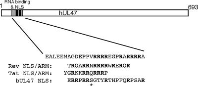
FIG. 9.
Schematic drawing of the hUL47 protein, showing the locations of the RNA binding domain and the NLS. The sequence of the minimal arginine-rich RNA binding motif is shown, together with the HIV-1 Rev and Tat ARMs and the bUL47 NLS. The asterisk denotes a serine residue in bUL47 conserved in several other UL47 homologues.

