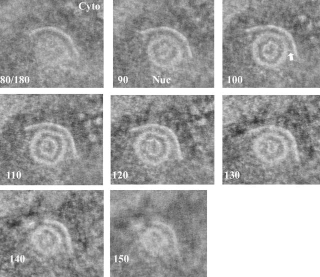
FIG. 1.
One-nanometer-thick slices from a tomographic reconstruction of a type B capsid attached to the INM. Cells infected with HSV-1 were high-pressure frozen and freeze substituted. The slice number of each image (out of a total of 180 slices) is indicated in the lower left of each panel. The cytoplasm is at the top of the image, and the nucleus is at the bottom. An arrow indicates a single bridging rod emanating obliquely from the capsid surface. Cyto, cytoplasm; Nuc, nucleus.

