FIG. 3.
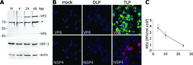
Analysis of IRF-3 and viral protein synthesis in RRV-treated mDCs. (A) mDC cultures were either mock-infected (M) or inoculated with RRV at an MOI of 5, and lysates were prepared at 4, 24, and 48 hpi and analyzed by Western blotting using the anti-RRV K230 serum (upper panel). The positions of the viral proteins and a murine protein reacting nonspecifically with the serum (*) are indicated at the right. The lysates were also probed with an anti-IRF-3 (middle panel) and an antiactin (lower panel) antibody. (B) mDC cultures were either mock infected or treated with TLP at an MOI of 5 or treated with an equivalent amount of DLP. Cells were processed for immunofluorescence at 24 hpi using anti-RRV VP6 (green) and anti-NSP4 (red). (C) The presence of newly produced infectious virus in RRV-treated mDC cultures was determined by titrating the virus supernatant collected at 4, 10, and 24 h. The data are shown as peroxidase-forming units (PFU)/ml of supernatant over time.

