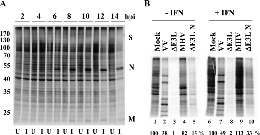
FIG. 2.
Protein synthesis in MHV A59-infected cells. (A) Time course of uninfected (U) and MHV infected (I) 17Cl1 cells. Cells were infected with WT virus at an MOI of 5. Cells were radiolabeled with [35S]methionine-cysteine for 30 min at the indicated times. Intracellular proteins were analyzed by SDS-PAGE and autoradiography. Positions of molecular weight markers in thousands (left) and of viral proteins (right) are indicated. (B) HeLa MHVR cells were mock pretreated or pretreated with IFN prior to infection with MHV, VV, VVΔE3L, or VVΔE3L N at an MOI of 5. Cells were pulse-labeled at 6 hpi for 30 min. Intracellular proteins were analyzed by SDS-PAGE and autoradiography. Proteins in each lane were quantified by densitometry and analyzed using ImageQuant software. Protein expression levels shown below each lane are expressed as the percentage of that measured for uninfected cells.

