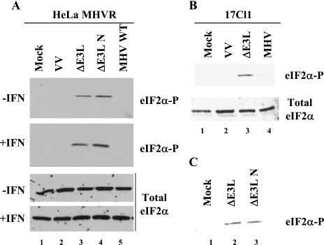
FIG. 3.
Detection of eIF2α phosphorylation in MHV A59- and VV-infected cells. HeLa MHVR (A) and mouse 17Cl1 (B) cells were infected with viruses as indicated above each lane. Cells were mock or IFN treated prior to infection. At 6 hpi cell lysates were analyzed for eIF2α by Western blotting with antibodies specific for the phosphorylated form of eIF2α and those that recognize both the phosphorylated and unphosphorylated forms (total eIF2α) of the protein. (C) Cell lysates from 17Cl1 cells infected with VVΔE3L or VVΔE3L N viruses were analyzed for eIF2α phosphorylation.

