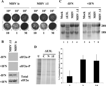
FIG. 7.
N protein is a type I IFN antagonist. (A) Monolayers of 17Cl1 cells were treated with increasing amounts (1 to 104 units) of type I IFN 24 h before infection. Cells were infected with the parental infectious clone-derived MHV A59 or ΔI virus at an MOI of 5. Plaques were stained with crystal violet at 48 hpi. (B) HeLa MHVR cells were mock or IFN treated prior to infection with MHV ΔI and WT MHV. At 6 hpi cell lysates were analyzed for eIF2α by Western blotting with antibodies specific for the phosphorylated form of eIF2α (two upper panels) and those that recognize both the phosphorylated and unphosphorylated forms of the protein for total eIF2α (lower two panels). The lanes shown were analyzed in parallel with results shown in Fig. 3A. (C) HeLa MHVR cells were infected with the viruses as indicated above each lane. Equal amounts of total RNA from cells at 6 hpi were analyzed by formaldehyde agarose gel electrophoresis and visualized by ethidium bromide staining. The positions of 18S and 28S rRNAs are indicated. (D) HeLa MHVR cells were transfected with either empty pCAGGS vector (C) or vector containing the WT N and ΔI N genes. Transfected cells were infected with VVΔE3L at 48 hpi and labeled 6 h later. Intracellular proteins were analyzed by SDS-PAGE and autoradiography. Proteins in each lane were quantified by densitometry. Measurements from three independent experiments were averaged. Error bars represent the standard deviations of the individual measurements.

