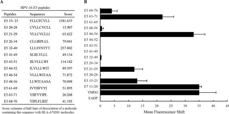
FIG. 1.
Identification of HLA-A*0201-restricted epitopes of HPV-16 E5 protein by T2 cell-binding assays. (A) Computer prediction of HPV-16 E5 peptides. (B) Stabilization of HLA-A*0201 molecules on the surfaces of T2 cells. T2 cells were incubated with peptides overnight at 26°C and then incubated at 37°C for 2 h, as described in Materials and Methods. HLA-A*0201 expression was determined by FACS staining with the monoclonal antibody PA2.1, and the mean fluorescence intensity (MFI) was calculated. The x axis indicates the mean fluorescence shift of T2 cells with tested peptide subtracted from that of T2 cells without peptide. Three experiments were conducted. The data are shown in the form of a histogram. YMDG, the HLA-A*0201 epitope of the tyrosinase-derived peptide YMDGTMSQV, which served as a positive control; E7 11-20, YMLDLQPETT, identified as an HLA-A2-specific CTL epitope of HPV-16 E7 protein, which also served as a positive control; EADP, the HLA-A1 binding peptide EADPTGHSY, which served as a negative control. The MFI values for the positive controls, YMDG and E7 11-20, were 36.8 ± 1.4 and 37.2 ± 7.6, respectively. The MFI value for the negative control, EADP, was 1.93 ± 0.7. The error bars indicate standard deviations.

