FIG. 4.
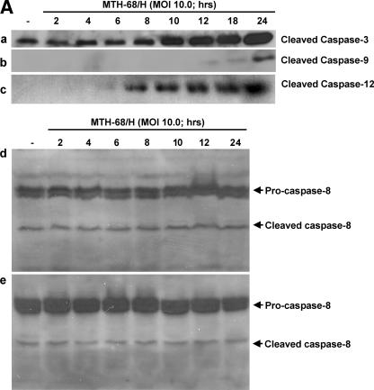
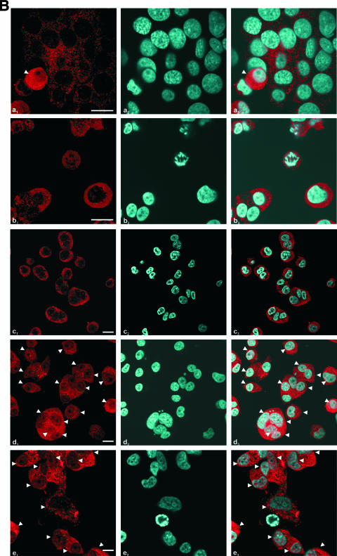
Analysis of the involvement of caspases in MTH-68/H-induced apoptosis. Cultures were infected with MTH-68/H as indicated in the figure. (A) Analysis of the cleavage of caspase-3, caspase-9, and caspase-12 upon MTH-68/H infection in PC12 cells (blots a, b, and c, respectively) and analysis of caspase-8 activation upon MTH-68/H infection in MCF-7 (blot d) and DU-145 (blot e) human cancer cell lines. (B) Analysis of nuclear translocation of ER stress-activated caspase-12 by immunocytochemistry in PC12 cells. The primary anti-caspase-12 antibody was detected by Cy3-conjugated goat anti-rabbit secondary antibody (micrographs a1 to e1), while nuclei were counterstained using Hoechst 33258 dye (micrographs a2 to e2). Micrographs a3 to e3 show the Cy3 and Hoechst 33258 channels merged. Micrograph a shows untreated cells, while b, c, d, and e show PC12 cells infected with MTH-68/H for 4, 6, 10, and 24 h, respectively. White arrowheads indicate cells with significant nuclear translocation of caspase-12. Scale bars, 15 μm.


