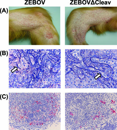
FIG. 1.
Comparison of monkeys infected with ZEBOV (left panels) or ZEBOVΔCleav (right panels). (A) Macular cutaneous rashes on day 6 postinfection. (B) Phosphotungstic acid hematoxylin-positive fibrin in spleen. Staining was carried out as described in reference 5. Note that there is no apparent difference in the amount or distribution of polymerized fibrin (see arrows that point to fibrin-stained regions). Original magnification, ×40. (C) Immunostaining of inguinal lymph nodes. Staining was carried out as described in reference 4. Note that positive immunostaining of monocytes-macrophages for Ebola virus (red) is evident in both animals. Also, lymphoid depletion and lymphocytolysis are prominent in both animals. Original magnification, ×20.

