FIG. 1.
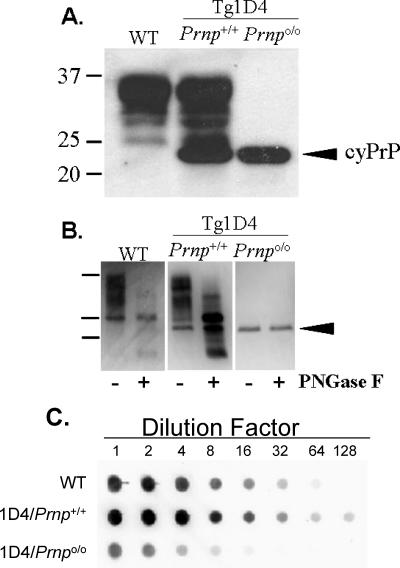
PrP characteristics in 1D4 mouse lines. A. Patterns of expression of PrPC and cyPrP from non-Tg (wild-type [WT]), Tg1D4(Prnp+/+), and Tg1D4(Prnpo/o) mice, detected by mouse-specific anti-PrP D13 antibody. Each lane represents ∼10 μg of total protein prepared from the brain of a representative mouse from each line. PrPC ranges from 25 to 35 kDa (lanes 1 and 2), representing the typical pattern of unglycosylated and glycosylated fractions, whereas cyPrP, because of the absence of the GPI anchor, migrates slightly faster, as a single ∼23-kDa band (lanes 2 and 3). This blot was overexposed to emphasize the banding patterns and may overestimate the level of cyPrP present. B. PrP from each mouse line was treated with PNGase F to confirm that cyPrP is not glycosylated. This treatment also allows a crude comparison of the relative levels of cyPrP and PrPC within the same brain of a Tg1D4(Prnp+/+) mouse. Densitometry estimated cyPrP to represent at ∼20 to 25% of PrPC. C. Serial dilutions of 1% brain homogenates prepared in lysis buffer from each mouse line were performed to more accurately assess the relative levels of PrPC and cyPrP. The image was processed on a Bio-Rad XRS document imager, and Quantity One (Bio-Rad) software was used to calculate the relative density against the dilution factor. This confirmed the expression level of cyPrP to be ∼20% that of wild-type mice. The higher total level of PrP in Tg1D4(Prnp+/+) mice reflects the combination of cyPrP and endogenous PrPC.

