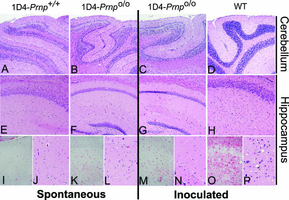
FIG. 4.
Pathological features of normal and Tg mice. Representative histological sections from the cerebellum (A to D) and hippocampus (E to P) of each experimental group, at low (A to H) and high (I to P) (×4) magnification, stained with hematoxylin and eosin (all except images in panels I, K, M, and O) to assess spongiform change or with anti-GFAP (I, K, M, and O) to assess gliosis within the hippocampus. Cerebellar granule cell layer atrophy was associated with cyPrP expression in both Tg1D4(Prnp+/+) and Tg1D4(Prnpo/o) mice, compared with age-matched wild-type (WT) mice. No vacuolation was evident in these mice at any level of magnification. When challenged with RML, Tg1D4(Prnpo/o) mice >500 days old did not show signs of vacuolation or gliosis, compared with the extensive vacuolation and gliosis present in 150-day-old WT mice that were symptomatic with clinical (ataxia, rough coat, hunched posture, and plastic tail) and biochemical (PK-resistant PrP) evidence of scrapie.

