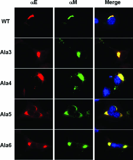
FIG. 5.
Colocalization of MHV E and M proteins. Mouse 17Cl1 cells were infected with wild-type (WT), Ala 3, Ala 4, Ala 5, and Ala 6 viruses. Cells were fixed and analyzed by immunofluorescence at 10 h p.i. using mouse and rabbit antibodies against the M and E proteins, respectively. Fluorescein isothiocyanate-conjugated mouse and AlexaFluor 594-conjugated rabbit secondary antibodies were used to visualize the localization of the proteins. Colocalization of M and E proteins is represented in the merged images by yellow. Nuclei were stained with DAPI.

