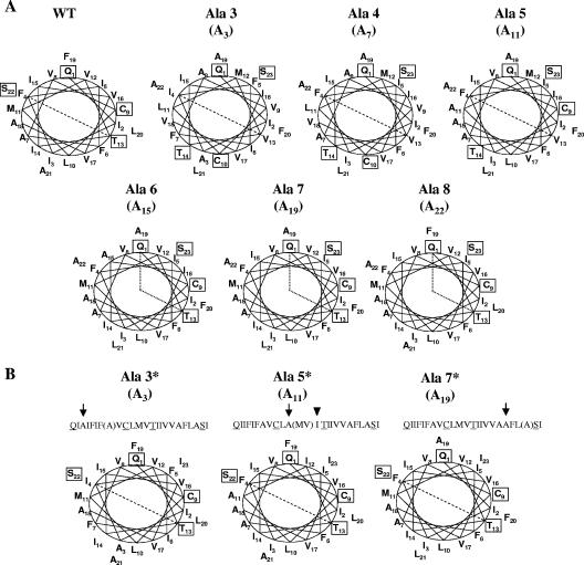
FIG. 6.
Helical wheel analysis of the E protein predicted transmembrane domains of alanine insertion mutant viruses. (A) Schematic top views from the amino ends of wild-type (WT) and alanine insertion mutant E protein transmembrane domains. Hydrophilic polar residues Q1, C9, T13, and S22, corresponding to Q15, C23, T27, and S36 in the full-length wild-type protein, are boxed. Residues toward the carboxy end of the helix are shown adjacent to the corresponding positions relative to the first 18 amino acids in the transmembrane domain. Numbers below the names indicate the position of the inserted alanine residue in the helical wheel. Lines across the wheels are added to emphasize the positioning on one face of the α-helix for wild-type, Ala 5*, Ala 3*, and Ala 7* viruses and the disruption of the positions of these residues in the Ala 3 to Ala 8 mutant proteins. Helical wheels plots were prepared using Gene Runner version 3.05. (B) Schematic tops views of Ala 3*, Ala 5*, and Ala 7* E proteins, illustrating positioning of the hydrophilic residues with removal of one amino acid on the carboxy-terminal side of the alanine insertion. Alanine insertions are indicated by arrows in the sequences above each wheel. Amino acids that were removed are enclosed in parentheses. The arrowhead notes the isoleucine insertion resulting from codon rearrangement in the recovered Ala 5* virus.

