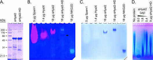
FIG. 1.
Characterization of sHyal2, sHyal2-HD, and HyalX. (A) SDS-PAGE analysis of sHyal2 and sHyal2-HD. (B) Native-PAGE zymography of HyalX, sHyal2, and sHyal2-HD. Pink clearings indicate hyaluronidase activity, and light-blue clearings are caused by protein. A faint pink band in the sHyal2-HD lane (arrow) likely represents residual HyalX activity. (C) Restaining of the gel shown in panel B with Coomassie blue reveals the positions of the protein species. Note that the amounts of Spam1 and HyalX listed above the gel refer to total protein of these relatively impure protein preparations, and the actual amounts of Spam1 and HyalX were below the limit of detection using Coomassie blue stain. (D) Hyaluronidase assay. Fifty-microgram samples of hyaluronan were incubated at 37°C at pH 5.6 in the presence of the indicated proteins for 14 h. The samples were then separated in a 0.5% agarose gel by electrophoresis and visualized with Stains-All.

