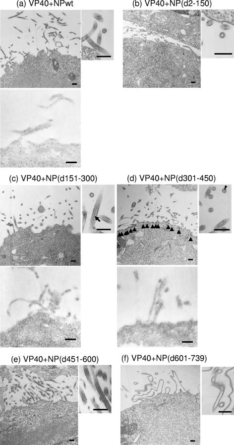
FIG. 5.
Electron microscopy of cells expressing VP40 and wild-type or mutant NPs. (a) Cell expressing VP40 and wild-type (wt) NP. NP helices were observed in VLPs budding from the cell surface as well as in the cytoplasm (upper panel). NPs within VLPs were immunolabeled with an anti-NP antibody conjugated with 5-nm gold particles (lower panel). (b) Cells expressing VP40 and NP(d2-150). NP helices were not found in the cytoplasm. Empty VLPs were released from cells. (c) Cells expressing VP40 and NP(d151-300). NP helices were not found in the cytoplasm, but electron-dense material (arrow), which reacted with an anti-NP antibody conjugated with 5-nm gold particles (lower panel), could be seen in VLPs. (d) Cells expressing VP40 and NP(d301-450). Small electron-dense material, which reacted with an anti-NP antibody conjugated with 5-nm gold particles (lower panel), was found at the plasma membrane (upper left panel, arrowheads) and in VLPs (upper right panel, arrowhead). (e) Cells expressing VP40 and NP(d451-600). NP helices were formed in the cytoplasm and incorporated into VLPs. (f) Cells expressing VP40 and NP(d601-739). NP helices were found in the cytoplasm but not in VLPs. Bars, 200 nm.

