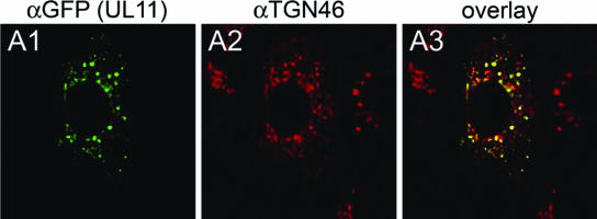
FIG. 6.
Digital images of confocal micrographs showing UL11 localization. Vero cell monolayers were transfected with the UL11-GFP-expressing plasmid. At 24 h posttransfection, cells were washed thoroughly, fixed, and stained with the anti-GFP antibody (A1) or with the Golgi specific marker TGN46 (A2) and fluorescence was visualized by confocal microscopy. A3, overlay. Magnification, ×63; zoom, ×2.

