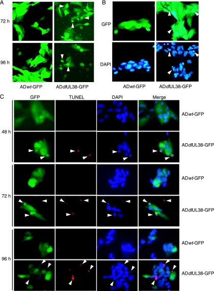
FIG. 6.
Cells infected with the pUL38 deletion mutant demonstrate morphological changes characteristic of apoptosis. (A) Normal HF cells were infected with ADwt-GFP or ADdlUL38-GFP at an input genome number equivalent to 1 PFU of wild-type virus/cell, and the morphology of infected GFP-positive cells was examined under a fluorescence microscope at 72 h and 96 h postinfection. Arrowheads indicate representative cell shrinkage, membrane blebbing, and vesicle release in ADdlUL38-GFP infection. (B) Infected cells were labeled with DAPI at 96 h postinfection. Condensed chromatin in cells infected with ADdlUL38-GFP is indicated by arrowheads. (C) Infected cells were colabeled with DAPI and TUNEL at the indicated times postinfection. The colocalization of GFP, TUNEL, and DAPI staining in the same ADdlUL38-GFP-infected cell population is indicated.

