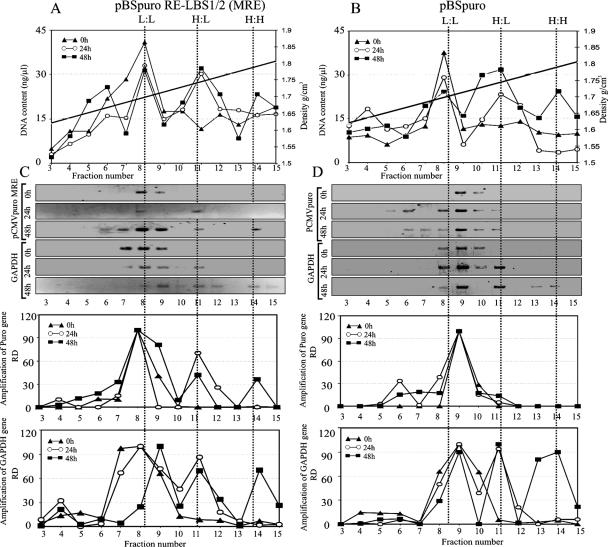
FIG. 2.
The plasmid containing the KSHV minimal replicator element replicates in synchrony with the cellular DNA. 293 cells cotransfected with pBSpuro MRE or pBSpuro and the LANA expression vector were labeled with 30 μg/ml BrdU for 0 h, 24 h, and 48 h, allowing no replication, one round, and two rounds of replication in the presence of the density label, respectively. DNA extracted from these cells was sonicated and separated on a CsCl density gradient. Fractions of 250 μl were collected from the top and subjected to density determination of each fraction and DNA extraction. Relative amounts of DNA in each fraction (fractions 3 to 15) are plotted against the densities of the fractions. (A and B) The distribution of total DNA from 293 cells transfected with pBSpuro MRE (A) or pBSpuro (B) in these fractions at 0 h (triangles), 24 h (circles), and 48 h (squares) is shown. Distribution of DNA was detected at three peaks with densities of 1.7, 1.75, and 1.8 g/cm3, which correspond to the unreplicated L:L, semireplicated H:L, and fully replicated H:H DNA molecules, respectively. Proportions of the transfected plasmid in these fractions were detected by PCR amplification using a specific primer targeted to the puromycin gene. Relative amounts of amplicons were quantified in these fractions. (C) Levels of pBSpuro MRE copies in these fractions (fractions 3 to 15) at 0 h (triangles), 24 h (circles), and 48 h (squares) after BrdU pulsing are presented based on the relative densities of the amplicons. Relative amounts of cellular DNA from these fractions, amplified using the GAPDH gene, were quantified, and the relative densities of the bands are presented as a graph. (D) Quantitation of the pBSpuro vector in different fractions of the gradient. Amplification of the puromycin target showed a peak of the plasmid template at only L:L density, suggesting no replication. Distribution of cellular DNA was detected as L:L, H:L, and H:H densities after 0, 24, and 48 h of BrdU pulsing, suggesting semiconservative replication of the cellular gene.

