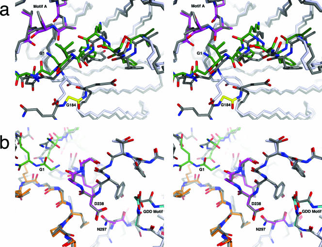
FIG. 5.
Comparison of the 3CD and 3Dpol structures at the N terminus, the active site, and the fingers domain shown in stereo. (a) The structure of the active site of the polymerase remains mostly unchanged between 3CD (light gray) and 3Dpol (dark gray). Notable residues of the 3Dpol active site are colored: Asp238, Ala239, Leu241, and Asn297 are magenta, the GDD motif (Asp328) is cyan, the N-terminal fingers are green, and the middle finger is orange. (b) One of the largest positional differences between 3CD and 3Dpol occurs within the fingers region of the 3D domain (3CD residues 226 to 251; 3Dpol residues 43 to 68). 3CD is dark gray with Gly184 in yellow; 3Dpol is light gray and green with Gly1 also in green and motif A in magenta. Note that the N-terminal G184 of 3CD is removed from the binding cleft, whereas Gly1 of 3Dpol is buried. Only slight differences are seen in motif A (residues 238 to 241 of 3Dpol), magenta in 3Dpol.

