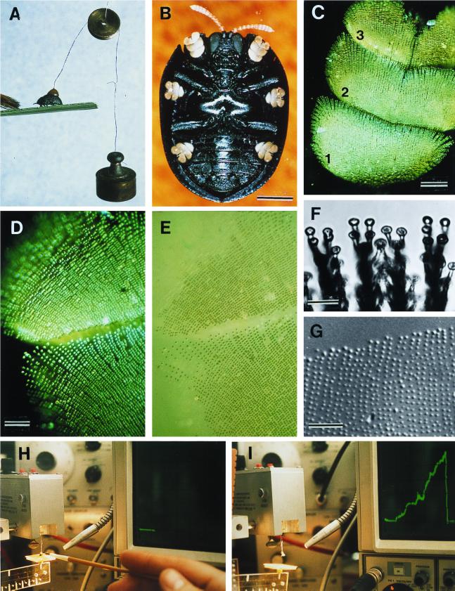
Figure 2.
(A) Beetle withstanding a 2-g pull; brush strokes are causing the beetle to adhere with its tarsi. (B) Ventral view of beetle, showing yellow tarsi. (C) Tarsus (numbers refer to tarsomeres). (D) Tarsus in contact with glass (polarized epi-illumination). (E) Same as preceding, in nonpolarized light; contact points of the bristles are seen to be wet. (F) Bristle pads, in contact with glass. (G) Droplets left on glass as part of a tarsal “footprint.” (H and I) Apparatus diagrammed in Fig. 1. In H, beetle is on platform, before lift is applied (horizontal trace on oscilloscope); in I, the lift has been applied (ascending green trace) to point where beetle has detached (return of trace to baseline). [Bars = 1 mm (B), 100 μm (C), 50 μm (D), 10 μm (F), and 50 μm (G).]

