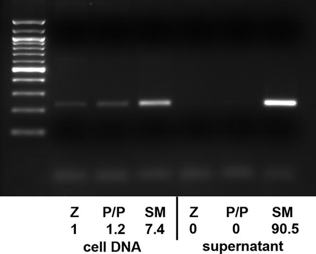
FIG. 5.

Detection of extracellular EBV virion DNA. SM-KO cells were transfected with Z alone (Z), Z plus BSLF1/BALF5 (P/P), or Z plus SM (SM). DNA was prepared from both cells and extracellular medium 72 h after transfection. Virion DNA was prepared from supernatant by DNase treatment followed by protease digestion prior to DNA isolation. PCR was performed with primers corresponding to the BamHI W fragment of EBV DNA, and products were analyzed by gel electrophoresis and staining with ethidium bromide. A 100-bp molecular size standard ladder is shown to the left. The same amount of DNA used for this analysis was also quantitated by quantitative PCR using a dye-labeled BamHI W probe. The relative amounts of EBV DNA as determined by the quantitative PCR are shown below each lane.
