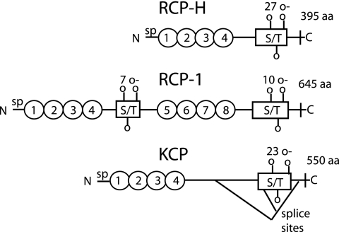
FIG. 2.
Analysis and comparison of predicted structures of RCP and KCP. All three proteins have a signal peptide (sp) in the N terminus. The ovals symbolize CCP domains. S/T boxes are areas with predicted O-glycosylated serine and threonine residues, where the number of O-glycosylation sites is indicated above the boxes. In the C terminus there is a transmembrane region followed by a single intracellular amino acid. Three isoforms of KCP have been identified due to splicing events, for which the two donor and the single acceptor sites have been identified (41).

