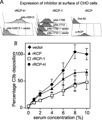
FIG. 7.
rRCP-H and rRCP-1 inhibit C3b deposition. CHO cells were transfected with constructs encoding either recombinant membrane-bound rRCP or rKCP. Negative control cells were transfected with an empty vector. (A) The levels of rRCP and rKCP expression by the transfected cells were determined by staining with polyclonal antibodies directed against CCP1-2 of RCP-H, CCP1-2 or CCP5-6 of RCP-1, and CCP1-2 of KCP. For rRCP expression, the negative controls were stained in the same manner as the rRCP-transfected cells, as indicated. For rKCP, the negative control is rKCP-expressing cells stained with only the secondary antibody. (B) C3b deposition was measured by flow cytometry. Cells were incubated with anti-hamster antibodies and different concentrations of human serum to activate complement and deposit C3b on the cell surface. The cells were then stained with an anti-C3b antibody. The experiment was performed twice in triplicate (or duplicate for rRCP-H). The geometric MFI obtained for the vector-only control cells in 10% serum was set as 100% C3b deposition. The C3b deposition percentages were calculated for all of the individual samples. The average and standard deviations of these values are plotted. A 100% C3b deposition corresponded to a geometric MFI of 195 or 125.

