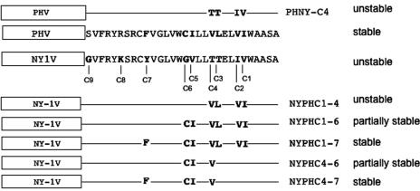
FIG. 6.
Schematic representation of NY-1V and PHV G1 tail mutants used in this study. The C-terminal 30 residues from NY-1V and PHV G1 tails were aligned using the ClustalW program, and differences at positions C1 to C9 are indicated (boldface). Mutations were incorporated into NY-1V (NYPH) or PHV (PHNY) as depicted, and a summary of mutant protein degradation by the proteasome is given on the right.

