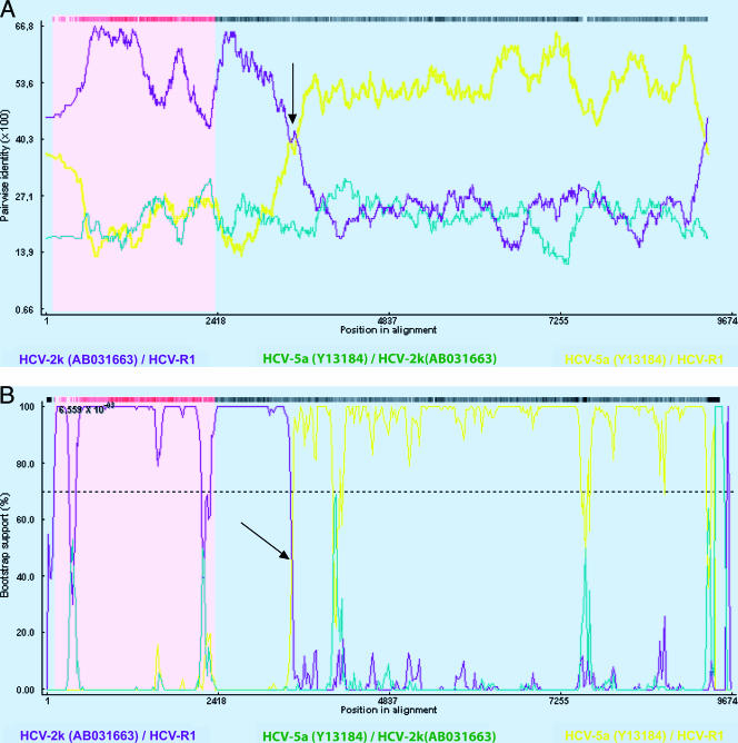
FIG. 3.
Determination of the recombination point with the Recombinant Detection Program method (A) or the BOOTSCAN method (B) using the Recombinant Detection Program version 2. Plots for comparing the R1 and 2k strains are shown in purple, and plots for comparing the R1 strain and the 5a reference strains are in yellow. An arrow indicates the crossover point. Reference sequences from HCV strains from the Los Alamos HCV sequence database are listed by their accession numbers.

