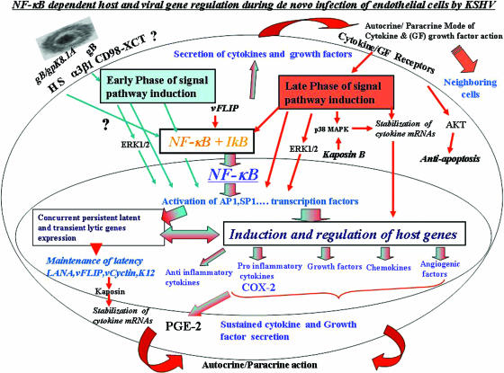
FIG. 10.
Schematic representation depicting the early and late induction phases of NF-κB during in vitro KSHV infection of HMVEC-d cells and their potential roles in transcription factor regulation, establishment and maintenance of KSHV infection, and cytokine secretion. In the early phase of NF-κB induction (blue arrows), virus binding and entry lead to signal pathway induction, such as FAK, Src, PI 3-K, AKT, PKC-ζ, MAPK-ERK1/2, and NF-κB signal molecules. Activated NF-κB translocates into the nucleus, which coincides with viral-DNA entry into the infected-cell nuclei, concurrent transient expression of limited viral lytic genes, and persistent latent gene expression. Overlapping with these events, a limited number of cytokines and growth factors are induced, which is initiated by transcription factors, like AP-1 (induced by ERK1/2 and NF-κB). Early activation of NF-κB and ERK1/2 also leads to the activation and release of NF-κB-inducible host factors, which act in autocrine and paracrine fashions on the infected, as well as neighboring, cells. The autocrine action of these factors, along with viral gene expression, probably contributes to the second, or late, phase of signal pathway activation (red arrows), including sustained NF-κB activation and phosphorylation of p38 MAPK, ERK1/2, and AKT required for the maintenance of latency. The blue and red arrows together indicate pathways induced during both early and late phases of KSHV infection.

