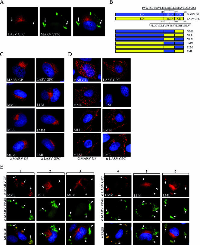
FIG. 2.
The transmembrane domain of MARV GP mediates accumulation of GP in MVBs upon coexpression with MARV VP40. HUH-7 cells were transfected with plasmids encoding VP40 and LASV GPC (A), chimeric glycoproteins (D), schematically presented in panel B, or chimeric glycoproteins and VP40 (E). Nuclei were counterstained with DAPI (4′,6′-diamidino-2-phenylindole). (A) HUH-7 cells were transfected with plasmids encoding LASV GPC and MARV VP40. At 24 h posttransfection, cells were fixed with 4% paraformaldehyde and immunostained with a rabbit anti-GPC and a monoclonal mouse anti-VP40 antibody. Bound antibodies were detected using secondary goat anti-rabbit IgG conjugated with rhodamine and goat anti-mouse IgG conjugated with FITC. The arrows show peripheral MARV VP40-positive clusters characterized as MVBs, which did not colocalize with LASV GPC. (B) Schematic presentation of MARV and LASV glycoproteins composed of the N-terminal ectodomain (ED), the hydrophobic membrane-spanning transmembrane domain (TMD) and the C-terminal cytoplasmic domain (CD). Chimeric proteins constructed by recombinant PCR are shown below and designated using a three letter code. The first letter of the construct's name indicates the origin of the ED (M, MARV; L, LASV), the second letter indicates the origin of the TMD, and the third letter indicates the origin of the CD. (C) Chimeric glycoproteins were expressed in HUH-7 cells and subjected to IF analysis as described above. Cells were immunostained with a rabbit anti-MARV GP or a rabbit anti-LASV GPC IgG depending on the origin of the ED. Bound antibodies were detected by a goat anti-rabbit antibody conjugated with rhodamine. (D) HUH-7 cells were transfected with plasmids encoding the chimeric glycoproteins and stained with goat anti-MARV IgG or rabbit anti-LASV GPC IgG at 4°C for 1 h. Cells were fixed but not permeabilized (note the difference from cells shown in panel C), and bound antibodies were detected using a donkey anti-goat IgG or a goat anti-rabbit IgG, both conjugated with rhodamine. (E) HUH-7 cells were cotransfected with plasmids encoding the chimeric glycoproteins and MARV VP40. At 24 h posttransfection, cells were fixed and immunostained with an anti-MARV VP40 monoclonal antibody and a rabbit anti-MARV GP (columns 1 to 3) or a rabbit anti-LASV GPC (columns 4 to 6) IgG. As secondary antibodies, a goat anti-mouse IgG conjugated with FITC and a goat anti-rabbit conjugated with rhodamine were used. Arrows indicate colocalization of VP40 and GP; arrowheads indicate VP40 located in peripheral MVBs showing no colocalization with expressed glycoproteins.

