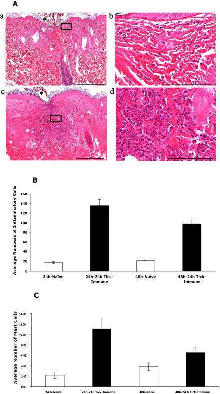
Figure 5. Histopathology of skin punch-biopsies from 24 h tick-immune animals shows increased inflammation.
Representative hematoxylin and eosin-stained sections of guinea pig skin punch biopsies obtained at 24 h near tick-attachment sites (*) from naive control (a, b), and 24 hour tick- immune (c, d). Samples from the 24-hour tick-immune animal had the highest number of inflammatory cells (c) which was characterized by a predominance of heterophils (d). In comparison inflammatory infiltrates were markedly reduced and comprised predominantly of mononuclear cells in 24-hour naïve animals (a, b). Scale bar = 500 µm (a, c); Scale bar = 100 µm (b, d). B. Dermal inflammatory cells in skin sections of 24 h tick-immune animals showed a statistically significant (P<0.001) increase in the numbers compared to naïve animals. C. Toluidine blue positive cells representing basophils/mast cells also showed a statistically significant increase (P<0.05) in skin biopsies of 24 h tick-immune animal compared to naïve animal (Error bars represent±SEM)

