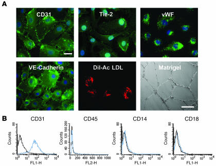
Figure 1. Phenotypic and functional characterization of EPCs.
(A) EPCs were fixed in 90% cold acetone and incubated with the appropriate primary antibody, then with a FITC-coupled secondary antibody. EPCs were characterized according to the presence or absence of endothelial-specific markers including CD31, Tie-2, vWF, vascular endothelial–cadherin (VE-cadherin), and uptake of Dil-acetylated LDL (Dil-Ac LDL) (scale bar: 10 μm) and by their capacity to induce tube formation on Matrigel (scale bar: 100 μm). (B) Flow cytometric analysis of surface antigens on EPCs. EPCs were positive for CD31 but not for monocytic markers CD45, CD14, and CD18 (blue histogram). Isotypic control is represented by the black histogram.

