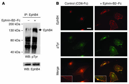
Figure 2. EphB4 activation by ephrin-B2–Fc.
(A) EPCs were stimulated with 3 μg/ml ephrin-B2–Fc for 30 minutes at 37°C. Cell lysates were prepared, subjected to immunoprecipitation with an anti-human EphB4 antibody, and resolved by SDS-PAGE, and proteins were transferred to nitrocellulose membranes as described in Methods. Membranes were then blotted with the 4G10 anti-phosphotyrosine antibody. To check for equal protein loading, membranes were stripped and reprobed with an anti-EphB4 antibody. pTyr, phosphotyrosine; WB, Western blot. (B) To confirm EphB4 activation, ephrin-B2–Fc–stimulated EPCs were subjected to double immunostaining with EphB4 and anti-phosphotyrosine antibodies. Note the membrane colocalization of the EphB4 and the 4G10 signals corresponding to clusters (yellow color) in ephrin-B2–stimulated cells. Inset: magnification showing clustering and internalization of the phosphorylated EphB4 when cells were stimulated with ephrin-B2–Fc (right) but not with control CD6-Fc (left). Scale bar: 10 μm.

