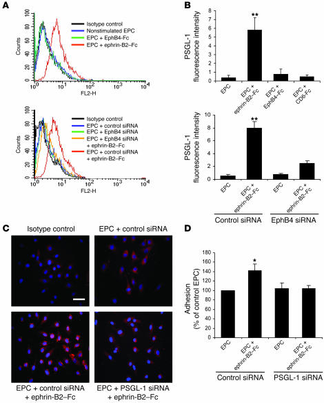
Figure 7. EphB4 activation mediates EPC adhesion through PSGL-1.
(A and B) PSGL-1 expression in EPCs. EPCs were stimulated with 3 μg/ml of either ephrin-B2–Fc, EphB4-Fc, or CD6-Fc and then processed for FACS analysis of PSGL-1 expression. (A) FACS profiles of nontransfected (top panel) and EphB4 siRNA–transfected (bottom panel) EPCs. (B) Expression of PSGL-1 in nontransfected (top panel) and EphB4 siRNA–transfected (bottom panel) EPCs. n = 3. **P < 0.01 versus nonstimulated EPCs. (C) Effect of PSGL-1 siRNA on PSGL-1 protein expression. Control siRNA– and PSGL-1 siRNA–transfected EPCs were stimulated with 3 μg/ml ephrin-B2–Fc or left unstimulated and then processed for immunocytochemistry with an anti–PSGL-1 antibody as described in Methods. PSGL-1–positive staining appears in red. Scale bar: 20 μm. (D) Effect of PSGL-1 siRNA on EPC adhesion to IL-1β prestimulated HUVEC monolayer. Adhesion was quantified by measuring OD at 570 nm. n = 3. *P < 0.05 versus nonstimulated EPCs transfected with control siRNA.

