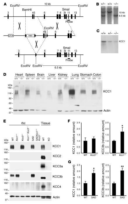
Figure 1. Generation of Kcc1–/– mice.
(A) Partial genomic organization of Slc12a4 (top) and the targeting construct (middle). Exons are shown as vertical bars, loxP sites as arrowheads. Transient Cre expression resulted in excision of exons 4 and 5 (bottom), producing a frameshift and premature stop. DTA, diphtheria toxin A cassette. (B) An additional EcoRV site was exploited for Southern blot analysis and resulted in the approximately 6.5-kb KO compared with the approximately 10-kb Kcc1+/+ fragment with the probe shown in A. (C) Northern blot analysis of Kcc1+/+, Kcc1+/–, and Kcc1–/– liver tissue with a full-length KCC1 probe revealed some residual aberrant transcript. (D) A membrane protein immunoblot with a KCC1 antibody confirmed the absence of KCC1 in tissues of Kcc1–/– mice. (E) rbc membrane protein immunoblot demonstrated KCC3b expression in WT rbc and absence of KCC3a, KCC2, and KCC4 in erythrocytes of WT, Kcc1–/–Kcc3–/–, and SADKcc1–/–Kcc3–/– mice. Actin served as a loading control. Kidney served as a positive control for KCC1 and KCC3b, brain for KCC2 and KCC3a, and lung for KCC4. (F and G) Expression levels of KCC1 and KCC3 proteins in rbc ghosts of various genotypes. (F) KCC3b protein level was upregulated in ghosts lacking KCC1, but KCC1 levels were unchanged in KCC3-KO ghosts. (G) Levels of both KCC1 and KCC3b proteins were increased in ghosts from SAD mice. Protein levels were determined by Western blot analysis (see Supplemental Figure 1) and normalized to actin. Bars represent arithmetic means from 6 mice normalized to WT. Error bars represent SEM. *P < 0.05, **P < 0.005 compared with WT.

