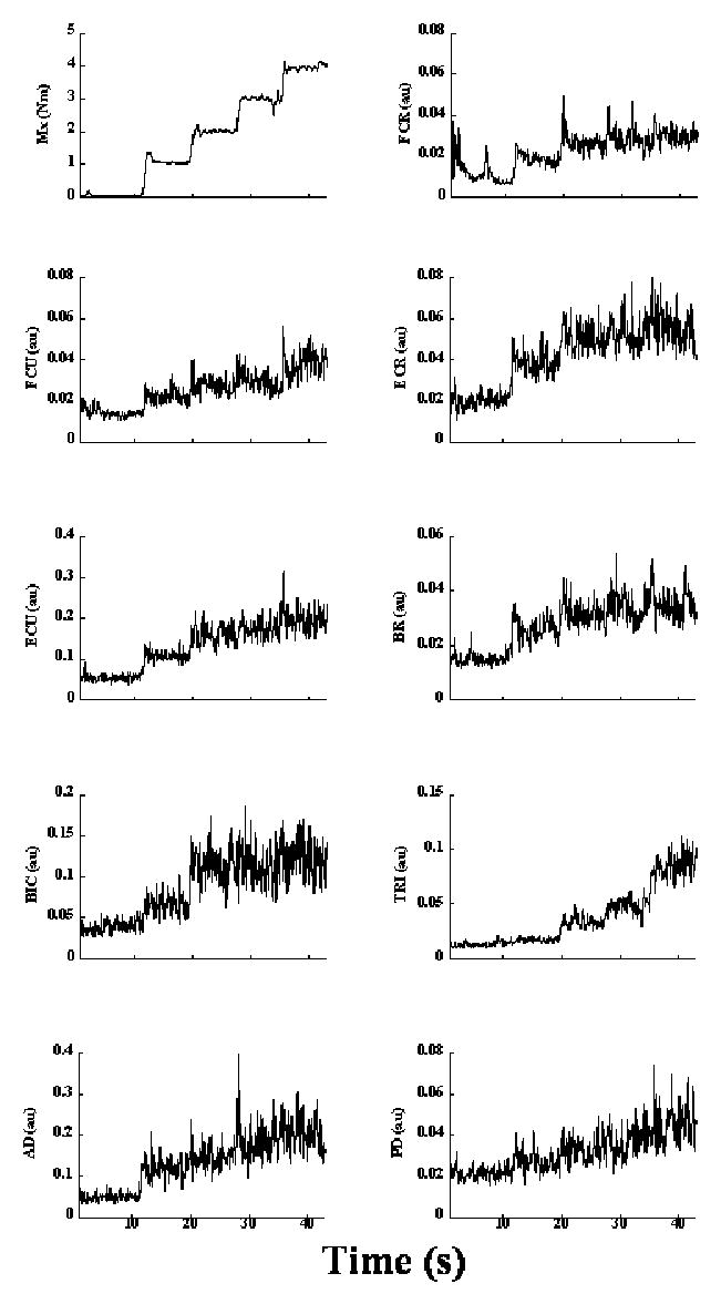
Figure 2. EMGs from a representative trial (subject s8) in the Step-push condition.

The top left panel shows the moment of force produced about the x-axis of transducer-1. All the remaining panels show corresponding EMG changes in the following muscles: flexor carpi radialis (FCR), flexor carpi ulnaris (FCU), extensor carpi radialis (ECR), extensor carpi ulnaris (ECU), brachioradialis (BR), biceps brachii (BIC), the medial head of triceps brachii (TRI), anterior deltoid (AD) and posterior deltoid (PD). The bursts in EMGs in some muscles (e.g. FCR) at the beginning of the trial correspond to the period when the arm was lifted to place the hand on the handle.
